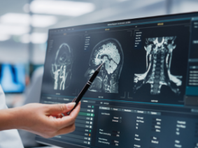
What is a Stereotactic Brain Biopsy?
Stereotactic brain biopsy is used to obtain tissue samples of suspicious growths or infections in the brain for more accurate diagnosis. It often is employed when lesions are located deep in the brain or when the patient cannot have general anesthesia.
Indications for a Brain Biopsy
After a suspicious growth or infection in the brain is found, the most common reasons a brain biopsy is required are:
- Tumors
- Abscess (infection)
- Inflammation (for example, encephalitis)
- Diseases of demyelination (for example, multiple sclerosis)
- Neurodegenerative disease (progressive degeneration of the brain, such as in Alzheimer’s disease)
A biopsy is useful in identifying suspicious areas or lesions that do not require surgery or to diagnose patients who are not able to have surgery, allowing them to seek other treatments for their condition.
Brain Biopsy Procedure
The stereotaxis procedure involves imaging tests, such as magnetic resonance imaging (MRI), computed tomography (CT) scans and three-dimensional computer imaging. These scans precisely target the area of the brain to be biopsied. This minimally- invasive-procedure requires a much smaller incision in the scalp than traditional craniotomy (open surgery), in which a piece of the skull is removed.
- The patient is placed under general anesthesia and the head safely secured.
- A small area of hair is shaved, and the incision site is clearly marked on the skin.
- Nickel-sized “fiducial markers” are gently stuck to different parts of the scalp, which provide reference landmarks for the stereotactic navigation system.
- A small hole is drilled into the skull.
- A needle is inserted through the small hole and guided by the imaging and computer software to the correct location.
- A tiny amount of brain tissue is taken out through the needle.
- The incision site is sewn up (stitches will need to be removed in 10-14 days).
- The tissue is sent to a pathologist, a doctor who determines the tumor’s type and grade or other diagnosis.
Stereotactic brain biopsy typically takes about 1-1/2 hours.
Risks of Brain Biopsy
Risks associated with the procedure are rare and include intracranial hemorrhage (bleeding inside the skull at the tissue site), infection and seizures. A failed attempt to obtain tissue for biopsy may require a repeat biopsy. This risk is also quite rare. Due to skilled neurosurgeons and state-of-the-art software and equipment, the procedure is very safe and crucial to an accurate diagnosis and tumor staging. There is also very little risk to the surrounding brain tissue.
Recovery and Follow-up
Following the procedure, bandages may be placed over the incision, which can be removed the next day. Patients are usually kept for observation before they go home and may need to stay in the hospital overnight as a precautionary measure for a specified time after the treatment before they go home, or they may be kept in the hospital overnight for observation.
Some patients do not experience any pain, though a bit of tenderness around the incision site is normal. Most patients can resume all their normal activities the next day.
Follow-up treatment will be decided based on the biopsy results. The neurosurgeon usually works with physicians specializing in radiation oncology and medical oncology (cancer) to develop a care and treatment plan. If the biopsy result is an infection, an infectious disease specialist will be called in.
Our neurosurgeons are experts in performing minimally invasive stereotactic brain biopsies. If you need a biopsy or have other concerning neurological symptoms, please contact us to make an appointment and discuss your concerns. Experience the care and compassion of the doctors and staff at Neurosurgical Associates of Central Jersey. Your care and comfort are our top priority.


