What Is a Chiari Malformation?
Chiari malformations (CMs) occur when brain tissue—the cerebellum—extends into the spinal canal through an opening in the base of the skull called the foramen magnum. It can be the result of an abnormally small or abnormally shaped skull. When the cerebellum goes through the narrow canal of the foramen magnum it puts pressure on it and the brainstem, which can affect the functions of these two areas of the brain and also restrict the flow of cerebrospinal fluid (CSF).
Chiari Malformation Causes and Risk Factors
There are three types of CMs. Type I can be a developmental form, occurring as the brain and skull are growing, but is more often congenital, meaning present at birth. These typically do not cause symptoms until adulthood. Types II and III are congenital and present during childhood. Congenital forms of CMs—also called primary CMs—are much more common than type I, or secondary, CMs. Primary CMs are due to structural defects in the brain and spinal cord. A genetic component may be involved, and maternal diet—a lack of certain vitamins and minerals—may also play a role.
Type II CMs, previously known as Arnold-Chiari malformations, almost always occur alongside another congenital condition known as spina bifida.
Secondary CMs happen when CSF leaks from the lumbar or thoracic spine. This is usually due to injury or infection.
Chiari Malformation Symptoms
Symptoms of Chiari malformations can include:
- Headaches, the most common symptom
- Syrinx or syringomyelia, a fluid-filled cyst that can compress the spinal cord
- Balance problems
- Hearing problems and tinnitus, a ringing or buzzing in the ears
- Hydrocephalus, an abnormal enlargement of the head that can affect infants with type II CMs, also known as Arnold-Chiari malformation
- Sleep apnea
- Difficulty swallowing
- Muscle weakness
- Scoliosis, an abnormal curvature of the spine
Chiari Malformation Diagnosis
A doctor may suspect a CM based on a medical history and physical exam, but imaging is needed to confirm. Imaging studies that can detect CMs include:
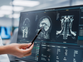
- Magnetic resonance imaging (MRI): MRIs use powerful magnets and radio waves to produce 3-D images of soft tissue, including brain tissue. It can provide images of the brainstem, cerebellum and spinal cord, which can show the presence of a CM.
- Computed tomography (CT): CT scans are multiple X-rays taken from different angles and combined with the help of a computer to produce cross-section images of the body. It can help detect brain abnormalities such as CMs.
Chiari Malformation Treatment
Often, people have CMs that do not cause symptoms. In this case, no treatment may be necessary at first. Neurologists and neurosurgeons will want to monitor those patients to make sure symptoms do not appear and the spinal cord does not become compressed.
Surgery is the treatment of choice for CMs that cause symptoms. The most common surgery is called a posterior fossa decompression, where the surgeon removes part of the back of the skull to relieve any pressure created by a CM or syrinx. People with hydrocephalus or a syrinx may need to have excess CSF drained.
If you suspect that you have a Chiari malformation of any type, request an appointment at Neurosurgical Associates of Central Jersey. Our surgeons have extensive experience treating CMs and will guide you every step of the way, from initial consultation to surgery to recovery and beyond.


